


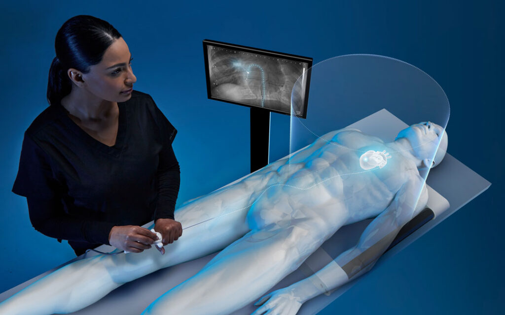
Electromagnetic Tracking for Cardiology
As Cardiology evolves across electrophysiology and catheter-based interventions, OEMs are integrating Electromagnetic (EM) tracking to support real-time device localization within moving cardiac anatomy. Aurora® EM tracking from NDI provides continuous 3D catheter localization data for OEM cardiology systems, complementing fluoroscopic workflows by providing positional data.
OEMs Shift from Imaging-Dependent Positioning to Continuous 3D Catheter Localization with Aurora®
Aurora provides the real-time localization layer that supports continuous catheter tracking, mapping accuracy, and reduced fluoroscopy dependence inside OEM-integrated cardiology workflows.
Continuous Localization
Enabling OEM Catheters to Track Throughout a Procedure. Aurora provides continuous 3D position and orientation data for catheter tips throughout the procedure. This enables OEM cardiology systems to maintain awareness of catheter position within moving cardiac anatomy without relying on intermittent fluoroscopic confirmation.
Mapping Accuracy
Supporting OEM Cardiology Platforms with Spatial Data for Mapping Workflows. Aurora supplies real-time positional data that OEM electroanatomical mapping systems can use to support the accuracy and density of cardiac maps. Continuous spatial data supports consistent map quality and reproducible catheter positioning during diagnostic and therapeutic procedures.
New Workflows
Opportunities for OEMs to Enable Low Fluoroscopy Workflows. Aurora provides continuous catheter localization data that OEMs may incorporate into system architectures. By providing continuous catheter localization independent of imaging, OEMs can design platforms that support lower-radiation and fluroless workflows across electrophysiology procedures.
Electromagnetic Tracking for Cardiology
Aurora integrates into OEM cardiology systems to provide continuous catheter localization across electrophysiology mapping, ablation, and catheter-based workflows.
Ablation Catheter
In OEM systems, EM tracked RFA/PFA catheters can be used to provide real time position and orientation within 3D electroanatomical mapping systems.
Mapping Catheter
EM tracked mapping catheters can be used to provide position data for localization of cardiac electrical activity as defined by the OEM.
ICE Catheter
In OEM systems, EM tracked ICE catheters can be used to support navigation within the heart as part of the larger mapping ecosystem.
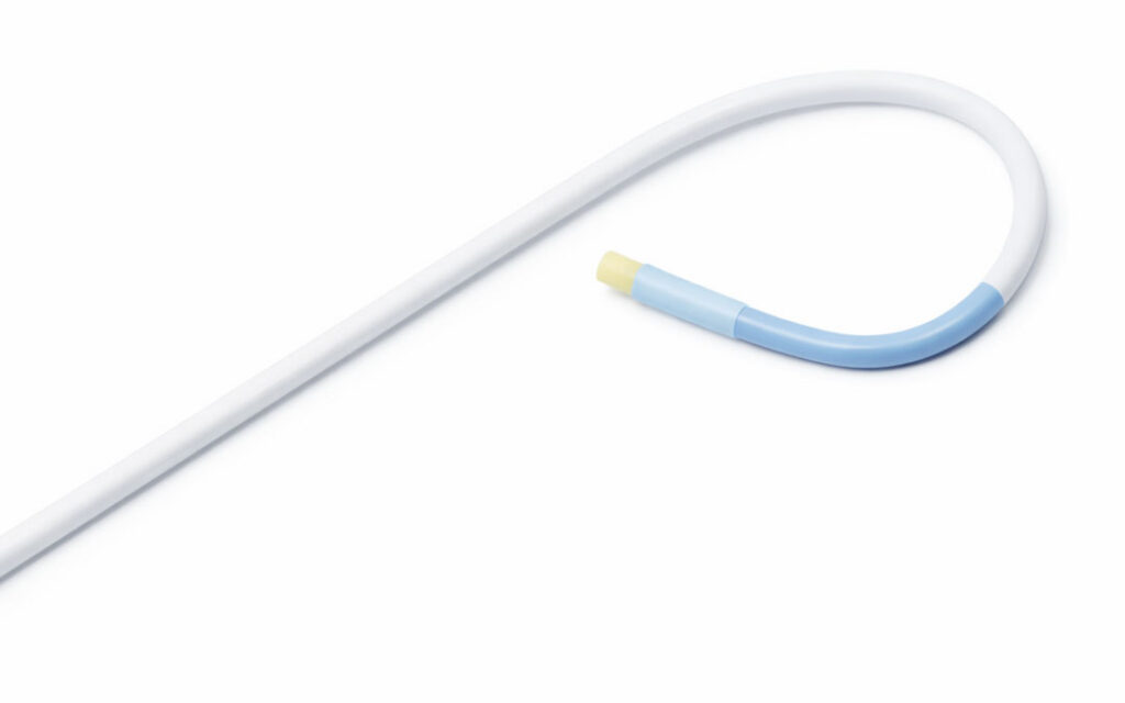
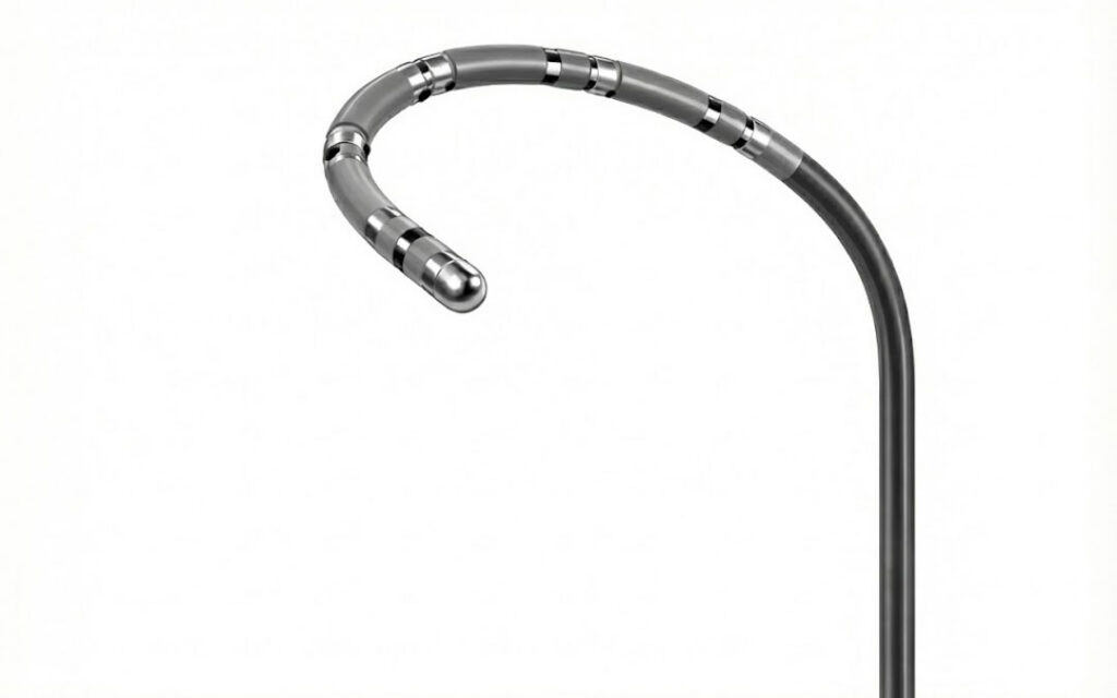
Steerable Sheaths
In OEM systems, EM tracked steerable sheaths can be used to support navigation to intricate cardiac structures.
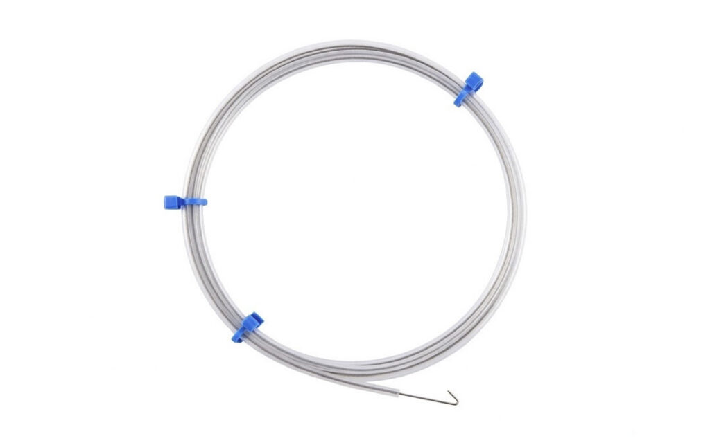
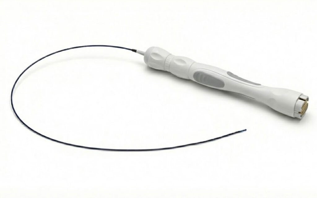
Guidewires
In OEM systems, EM tracked guidewires can be used to support 3D navigation in minimally invasive procedures in the heart.
Representative OEM Devices
Examples shown are representative of common OEM integrations.
Recommended Products
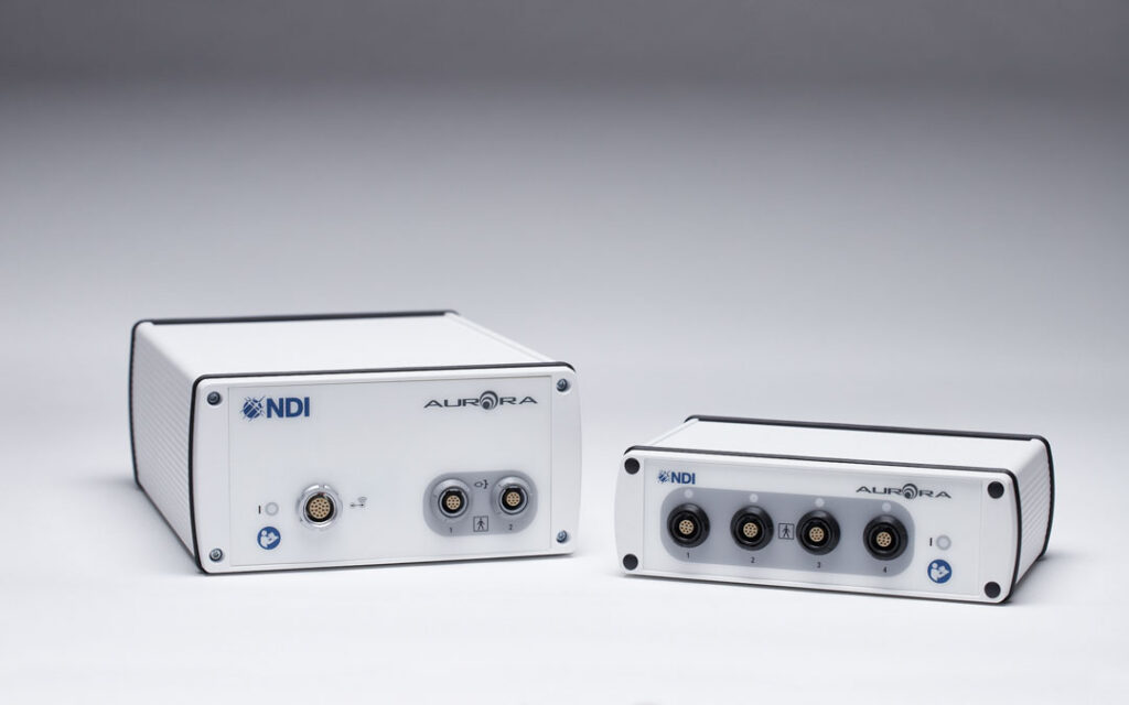
Aurora Electronics Units
Designed for integration into systems operating in complex C-arm workflows, offering high accuracy and a versatile range of configuration options.
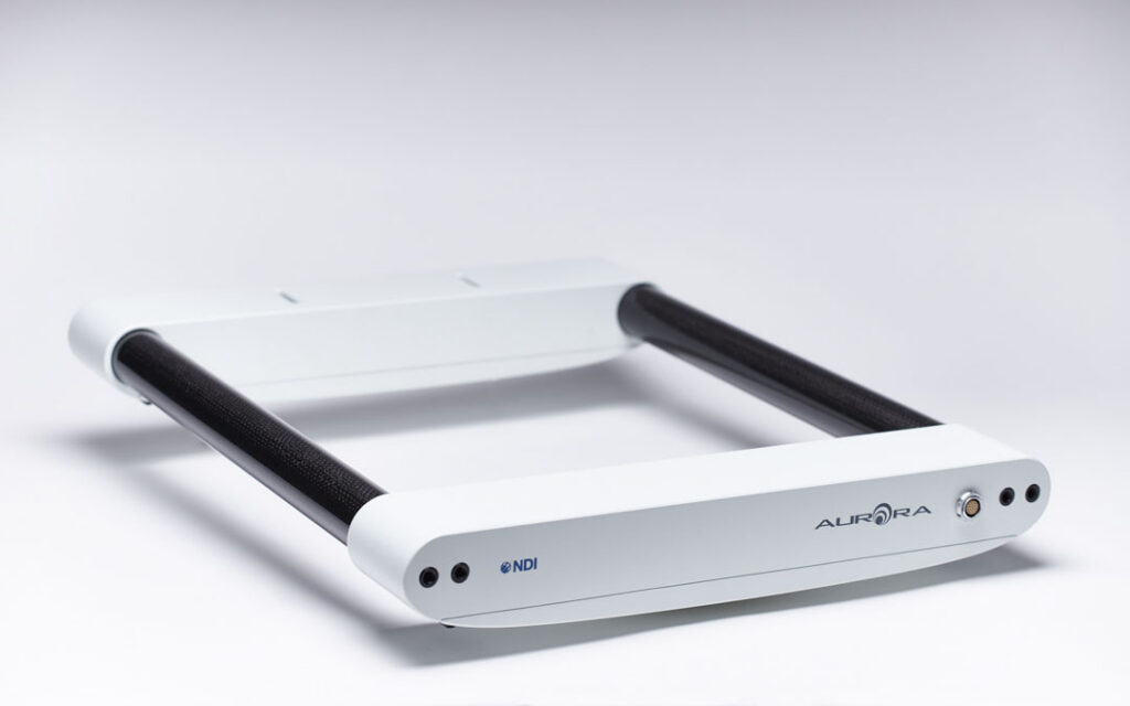
Aurora Window 50-60 Field Generator
Mounts under the patient table and out of sight during C-arm imaging. Accurate tracking is maintained without the introduction of imaging artefacts.
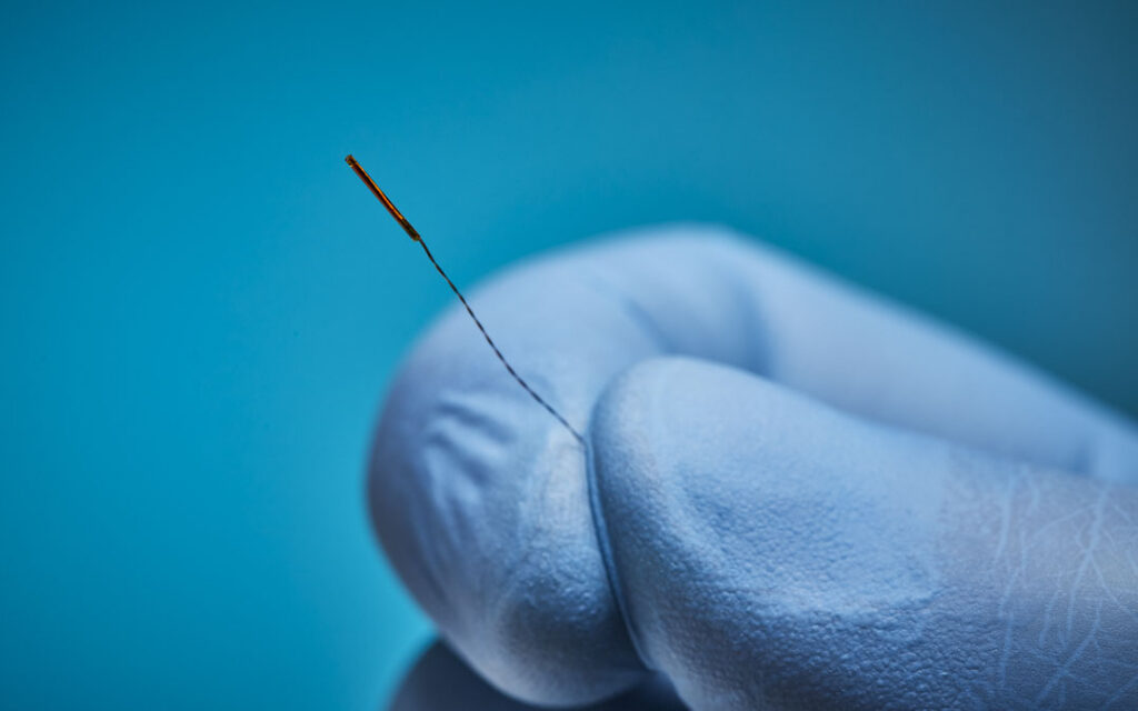
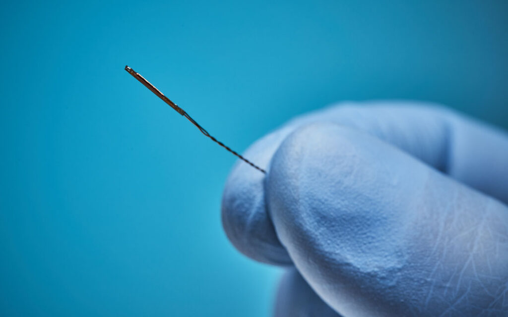
Aurora Sensors
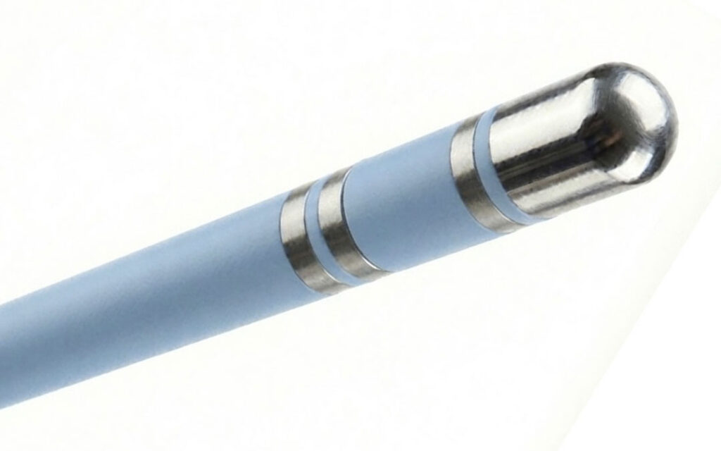
Ablation Catheter Sensor
Popular Choice: 5DOF Solid Sensor
Withstands high-voltage ablation environments while maintaining accurate tracking.
Mapping Catheter Sensor
Popular Choice: 5DOF Solid Sensor
Small sensors are critical for integration into various locations within the mapping catheter. 5DOF provides the smallest sensors.
Ice Catheter Sensor
Popular Choice: 6DOF Solid Sensor
Ice catheters require 6DOF tracking to enable image fusion of a preoperative CT scan
Steerable Sheath Sensor
Popular Choice: 5DOF Solid Sensor
Its small size allows for more flexibility in narrow anatomy
Guidewire Sensor
Popular Choice: 5DOF Solid Sensor
Its small size allows for more flexibility in narrow anatomy
Developing a Next-Generation Cardiology Platform?
Connect with our Product Integration team to identify the right Aurora tracking configuration for your electrophysiology, structural heart, or catheter navigation system.
Frequently Asked Questions: Electromagnetic Tracking for Cardiology
Why does tracking matter in cardiology?
Cardiology procedures across electrophysiology and structural heart interventions require precise catheter positioning inside the beating heart for diagnostic mapping, ablation therapy, and interventional access. Current workflows rely on a combination of fluoroscopy and electroanatomical mapping systems, but these approaches have limitations: fluoroscopy provides only 2D visualization with radiation exposure, and mapping systems depend on stable positional accuracy despite continuous cardiac motion. Electromagnetic tracking addresses these limitations by providing continuous, real-time 3D catheter localization. For OEMs developing cardiology platforms, EM tracking provides data that supports: precise catheter tip position and orientation tracking, improved mapping system accuracy through continuous spatial data, multi-catheter tracking during complex procedures, and reduced dependence on fluoroscopy. As cardiology procedures grow in complexity with next-generation ablation technologies such as pulsed field ablation (PFA) and expanding structural heart applications, continuous catheter tracking is becoming a foundational capability in cardiology system design.
What types of cardiology procedures does NDI electromagnetic tracking support?
NDI Aurora supports OEM development of cardiology systems for cardiac mapping, ablation (including pulsed field ablation), transseptal access, intracardiac echocardiography (ICE) integration, and structural heart navigation. Aurora provides continuous 3D catheter localization inside the beating heart across both electrophysiology and structural heart workflows.
How does Aurora support pulsed field ablation (PFA) workflows?
Aurora supports multi-sensor tracking on PFA catheters, providing positioning data that supports catheter placement workflows within OEM-integrated cardiology systems. The system can track up to 32 sensors with 5DOF and 16 sensors with 6DOF, accommodating the multi-sensor catheter designs used in next-generation PFA platforms.
Which field generator is recommended for cardiology procedures?
The Window 50-60 Field Generator is recommended for cardiology workflows. It is placed beneath the patient table, keeping the clinical workspace clear and avoiding interference with C-arm imaging. The Window Field Generator maintains accurate tracking without introducing imaging artefacts during fluoroscopy-based procedures.
What is the smallest sensor NDI offers for cardiology catheters?
Aurora supports sensors with diameters as small as 0.3 mm, enabling integration into ablation catheters, mapping catheters, diagnostic catheters, ICE catheters, steerable sheaths, and guidewires used across electrophysiology and structural heart procedures.
Can Aurora track multiple catheters simultaneously?
Yes. Aurora supports simultaneous tracking of multiple sensors, which is essential for cardiology procedures that involve mapping, ablation, and diagnostic catheters positioned concurrently inside the heart.
How does electromagnetic tracking reduce fluoroscopy dependence in cardiology?
Aurora provides continuous 3D positional data that OEM cardiology systems can incorporate into their catheter tracking workflows. OEM systems that integrate EM tracking may design reduced-fluoroscopy or fluoroless workflow architectures, subject to the OEM system’s regulatory clearance and clinical validation..